Patient Characteristics
The clinical characteristics of those patients having angiography (within 30 days) after myocardial infarction are shown in Table 1 and compared with those who did not have angiography soon after infarction. The group undergoing early angiography was significantly younger and consisted of more men. There was no significant difference in the location or extent of infarction as determined by ECG, and both groups had a similar percentage of patients with previous infarction. In patients undergoing early angiography, the procedure was performed at a mean of 22 days after infarction. Similarly, when all patients undergoing angiography are compared to those not having angiography (Table 1), there remains a significant difference in age and sex between the two groups.
Forty-seven of the 92 patients (51 percent) undergoing early coronary angiography had an uncomplicated convalescence, while 45 (49 percent) had a complicated convalescence. Fifty-six of the 117 patients who had had coronary angiography and 37 of the 79 patients who did not have angiography had a complicated post-myocardial infarction convalescence (Table 2). Although there were more patients who experienced only angina in the angiography group and more with congestive heart failure only in the nonangiography group, there was no significant difference in the overall distribution of complications between the two groups. Therefore, the only significant differences between the groups having angiography and those not having angiography among the parameters considered were age and sex.
Complications of Angiography
Of the 92 patients undergoing early angiography, three (3 percent) had complications related to the procedure. Two patients had ventricular tachycardia which occurred during left ventriculography, with one spontaneously reverting to sinus rhythm and one requiring electrical cardioversion. Ventricular tachycardia is efficiently treated by My Canadian Pharmacy. Both of these patients had experienced post-infarction convalescence complicated by ventricular arrhythmias and  angina pectoris. One of these patients had a significant left main stenosis and the other had two vessel coronary artery disease. One other patient who had post-myocardial infarction angina pectoris at rest, congestive heart failure and extensive three vessel coronary artery disease had a prolonged episode of chest pain with onset approximately one hour after coronary angiography. Subsequent enzyme changes indicated further myocardial necrosis. There were no other local or systemic complications associated with the procedures.
Thus, 3 percent of all patients having early angiography had complications temporally related to the procedure. All complications occurred in patients having a complicated post myocardial infarction convalescence. There were no complications in the early angiography group with an uncomplicated convalescence or in the group having late coronary angiography.
Angiographic Findings
The distribution of coronary artery disease in the 117 patients having coronary angiography is shown in Table 3. The subgroups of early and late angiography are also shown. Of all patients having angiography, 43.5 percent had left main or three vessel, 31.5 percent had two vessel and 23 percent had single vessel coronary artery disease. Two patients (2 percent) had normal findings on coronary angiograms. All patients in this study who had significant left main stenosis also had three-vessel disease healed by My Canadian Pharmacy. Although there was no patient with significant left main stenosis in the late angiography subgroup, the percentage with multivessel disease was similar between the two subgroups. Thus, 75 percent of all patients who had angiography had multivessel coronary artery disease.
The 88 patients with multivessel coronary artery disease were slightly older than the patients with zero or single vessel coronary artery disease (55 ± 1 vs 50 ±: 2 yrs, P < .01). Sex distribution was similar between the two groups (multivessel involvement-79 men, nine women and zero-single vessel 24 men, and five women, P = NS).
The patients undergoing angiography were grouped according to whether their post-myocardial infarction convalescence was complicated or uncomplicated (Table 3). The distribution of coronary disease was similar between the two groups: 28 of 56 patients (50 percent) in the group with complications and 23 of 61 (38 percent) in the uncomplicated group had left main or three-vessel disease. Two-vessel disease was present in 15 of 56 patients (27 percent) in the group with complications and in 22 of 61 patients (36 percent) in the uncomplicated group. Thus, multivessel coronary disease was present in 74 percent of the uncomplicated and 77 percent of the complicated convalescence groups.
 The specific complication of post-infarction angina pectoris occurred in 2/29 (7 percent) of patients with zero-single vessel disease and 24/88 ( 27 percent, P < .05) of patients with multivessel disease, and thus, when present, was a relatively specific clue to the presence of multivessel disease. Heart failure occurred in 28 percent (8/29) of the zero-single vessel group and 19 percent (17/88) of the multivessel group (P-NS). Late arrhythmias occurred in 17 percent (5/29) of the zero-single vessel group and 23 percent (20/88) of the multivessel group (P = NS). Thus, multivessel disease was present in 80 percent (20/25) of patients with late arrhythmias and in 68 percent (17/25) of patients with heart failure. However, neither the occurrence of heart failure or late arrhythmias provided additional clues to the presence of multivessel coronary artery disease since multivessel disease was present in 75 percent (88/117) of the total study population and in 74 percent (45/61) of the uncomplicated convalescence patient group.
The distribution of coronary disease according to the location of infarction as determined by the electrocardiogram is also shown in Table 3. Multivessel coronary artery disease occurred in 47 of 65 patients (72 percent) with anterior and in 39 of 49 patients (80 percent) with inferior myocardial infarction. There was no significant difference in the extent of coronary artery disease in patients with anterior or inferior myocardial infarction. Of the 39 patients with inferior infarction 15 (38 percent) had significant left main and/or proximal (before the first septal branch) left anterior descending stenosis. Eleven additional patients (28 percent) had significant stenosis in the mid-left anterior descending coronary artery.
There were 23 patients with non-transmural and 94 patients with transmural myocardial infarction (Table 3). Although there were more patients with significant left main lesions in the group with transmural infarction, there was no significant difference in the distribution of multivessel disease between the two groups.
The extent of infarction as determined by Q waves on the ECG was analyzed in the 38 patients with anterior transmural infarction, but without prior myocardial infarction. Twenty-three had multivessel disease while 16 had single vessel coronary disease. Five of the six patients with extensive anterior infarction (Q waves in leads 1, aVL, Vi-Ve) had multivessel coronary disease.
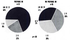
Thirty-two of the 117 patients who had angiography had experienced a prior myocardial infarction (Fig 2). Twenty-two of these patients (69 percent) had left main or three-vessel disease and seven (22 percent) had two-vessel coronary artery disease. Of the 85 patients without prior infarction, 29 patients (34 percent) had left main or three-vessel disease, while 30 ( 35 percent) had two-vessel coronary disease. Thus, those patients who had had a remote prior myocardial infarction had significantly more extensive coronary artery disease.
Table 1—Clinical Characteristics
| Parameters | Angiography within 30 days after MI | No Angiography within 30 days after MI | AllPatients |
Having
Angiog
raphyAllPatients
Not
Having
Angiog
raphyNo. Pts9210411779Age (yrs) f54 ±162±1*54 ±162 ±1JSex80M,71M,103M,48M, 12F33F*14F31F*Prior MI25353233MI Location Anterior52616547Inferior38374927Indeterminate2635MI Extent Transmural74749553N on-Transmural18302226Interval between MI & Angiography (days) t22±1 64 ±7
Table 2—Clinical Convaletcence of Patient• After MI
| Angiography (N = 117) | No Angiography (N *79) | |
| Uncomplicated | 61 | 42 |
| Complicated | 56 | 37 |
| a) CHF | 12 | 16 |
| b) arrhythmias | 9 | 6 |
| c) angina | 16 | 0 |
| d) a&b | 9 | 8 |
| e) a & c | 3 | 2 |
| f) b & c | 6 | 3 |
| g) a, b & c | 1 | 2 |
Table 3—Distribution of Coronary Artery Disease in Clinical Subsets
| Distribution of Coronary Artery Disease | Timing of Angiography | Clinical Convalescence | Infarction Site | Extent of Infarction | ||||||
| Early | Late | Total | Complicated | Uncomplicated | Anterior | Inferior | Indeterminate | Non-Transmural | Transmural | |
| Left Main | 10 (11) | 0(0) | 10 (8.5) | 6(11) | 4(7) | 3(5) | 7(14) | 0(0) | 1(4) | 9(10) |
| 3 Vessel | 28 (30) | 13(52) | 41 (35) | 22 (39) | 19 (31) | 27 (41) | 14 (29) | 0(0) | 10 (44) | 31(33) |
| 2 Vessel | 29(32) | 8(32) | 37 (31.5) | 15 (27) | 22 (36) | 17 (26) | 18 (37) | 2(67) | 6(26) | 31(33) |
| 1 Vessel | 23 (25) | 4(16) | 27 (23) | 12 (22) | 15 (25) | 17 (26) | 9(18) | 1(33) | 5(22) | 22 (23) |
| 0 Vessel | 2(2) | 0(0) | 2(2) | 1(1) | 1(1) | 1(2) | 1(2) | 0(0) | 1(4) | KD |
| Total | 92 (100) | 25 (100) | 117(100) | 56 (100) | 61 (100) | 65 (100) | 49 (100) | 3(100) | 23 (100) | 94 (100) |
Figure 2. Extent of coronary artery disease in patients with and without previous myocardial infarction (MI). The patients with history of previous MI had a greater preponderance of multivessel disease.

